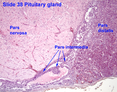
Pars intermedia
From Infogalactic: the planetary knowledge core
| Pars intermedia | |
|---|---|
| File:Gray1181.png
Median sagittal through the hypophysis of an adult monkey. (Pars intermedia labeled at bottom center.)
|
|
| Details | |
| Latin | pars intermedia adenohypophyseos |
| Identifiers | |
| Code | TH H3.08.02.2.00007 |
| Dorlands /Elsevier |
p_07/12616939 |
| TA | Lua error in Module:Wikidata at line 247: invalid escape sequence near '"^'. |
| TH | {{#property:P1694}} |
| TE | {{#property:P1693}} |
| FMA | {{#property:P1402}} |
| Anatomical terminology
[[[d:Lua error in Module:Wikidata at line 247: invalid escape sequence near '"^'.|edit on Wikidata]]]
|
|
Pars intermedia is the boundary between the anterior and posterior lobes of the pituitary. It contains three types of cells - basophils, chromophobes, and colloid-filled cysts. The cysts are the remainder of Rathke’s pouch.
In human fetal life, this area produces melanocyte stimulating hormone or MSH which causes the release of melanin pigment in skin melanocytes (pigment cells). However, the pars intermedia is normally either very small or entirely absent in adulthood.
In lower vertebrates (fish, amphibians) MSH from the pars intermedia is responsible for darkening of the skin, often in response to changes in background color. This color change is due to MSH stimulating the dispersion of melanin pigment in dermal (skin) melanophore cells.
External links
- Histology image: 14001loa – Histology Learning System at Boston University
- Histology image: 14101loa – Histology Learning System at Boston University
- Histology image: 38_11 at the University of Oklahoma Health Sciences Center
- UIUC Histology Subject 991